
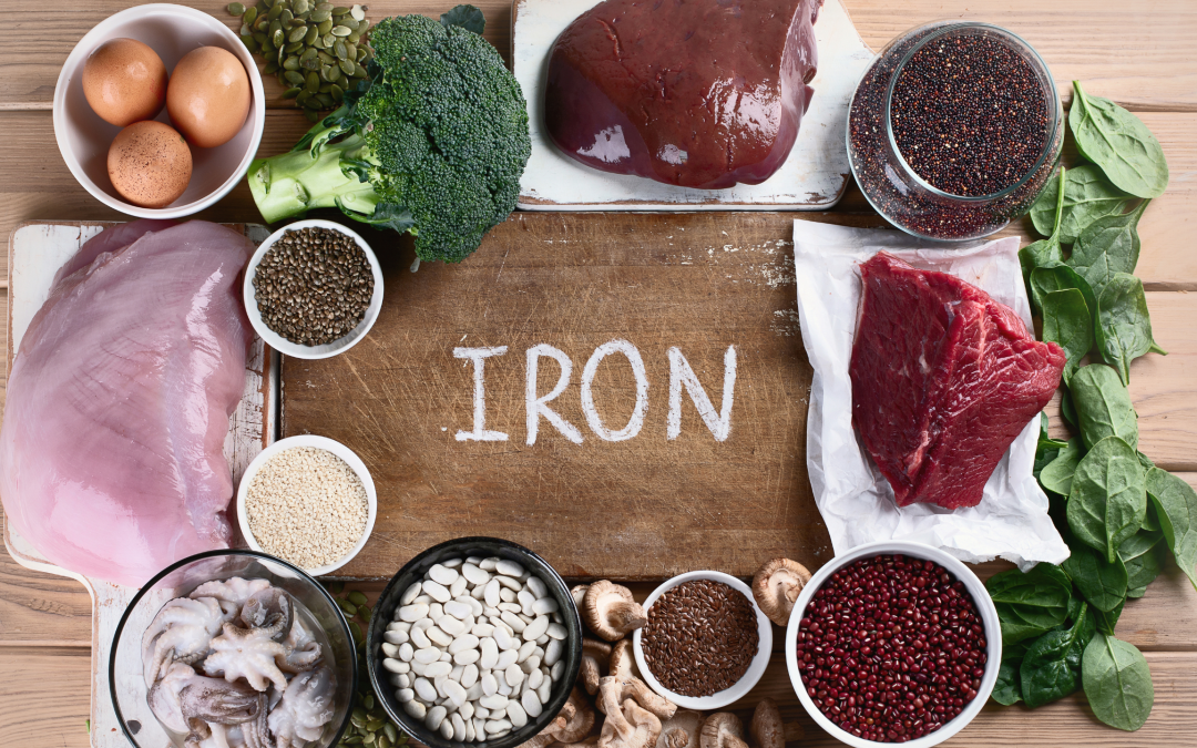
Over the past few years of working exclusively with IC patients, I have run across a very high percentage of my patients have oxidative stress and excessive amounts of reactive oxygen species. In addition, there is a very high percentage of them with Copper dysregulation and disorders in iron metabolism. This can be presented as both iron deficiency or iron overload, and there are certainly genetics that play a role such as those associated with hemochromatosis (HFE).
In this paper, I want to describe a mechanism of induced oxidative damage called Ferroptosis.
In part 2, I will discuss some therapies to deal with iron dysregulation if Ferroptosis is suspected.
Ferroptosis
Cell metabolism is precisely controlled for the maintenance of intracellular homeostasis. ATP is the major energy source for cell growth. The progressive oxidation of ingested nutrients provides electrons, and the continuous transfer of electrons in the ETC (electron transport chain) allows the cells to produce ATP continuously and steadily. However, mitochondrial ATP production is accompanied by the generation of reactive oxygen species (ROS), which is the normal by-product of aerobic metabolism.
The excessive reactive oxygen species (ROS) accumulation leads to oxidative stress and can, in combination with free ferrous iron (Fe2+), lead to ferroptosis.
What is ferroptosis?
It is a new entity discovered in 2012. The word is derived from ferro meaning “iron” and “ptosis” denoting falling. It is a new form of non-programmed cell death.
Ferroptosis is induced by the overproduction of phospholipid hydroperoxides, which is different from apoptosis, necrosis, and autophagy.
The basic premise revolves around lipid peroxidation.
The hallmark event of ferroptosis is the inactivation of the glutathione peroxidase 4 (GPX4), the subsequent accumulation of ROS, and, in combination with free ferrous iron (Fe2+), the increased damage to the membrane lipids.
It can cause oxidative damage to cell membranes- leading to cell death. This is due to increased intracellular iron that leads to increased reactive oxygen species. It is characterized by iron accumulation and lipid peroxidation as important markers of oxidative damage. This overwhelms the glutathione dependent antioxidant mechanisms leading to unchecked membrane lipid peroxidation, leading to loss of membrane permeability. This is what leads to cell death.
It is an iron dependent, non-apoptotic form of cell death. It plays an important role of development of various inflammatory related diseases.
This video below is very helpful to understand this process in a cartoon format that may be easier to digest
Below is a quick summary of this pathway.

In simple terms here is the general pathway.
Oxygen comes in through NOX enzyme. The NOX enzyme is upregulated by environmental factors (some people call this the NADPH steal)- creating superoxide through the superoxide dismutase enzyme (SOD). When this happens, we get hydrogen peroxide- and then when it combines with iron (through the FENTON REACTION)-it generates hydroxyl radicals.
If we don’t use our fats properly and through ALOX enzymes- we make Phospholipid Hydroperoxide. We need GPX4, coq10 and BH4 to calm this down. Dopamine can also inhibit this process too.
There are many reasons why you may be low in these elements.
For example, if there are too many free radicals, your reduced glutathione may be depleted trying to deal with it, not leaving enough of it to support the GPX4 enzyme, which relies on reduced glutathione. Or someone people are weak in the glutathione recycling, and their oxidized glutathione does not properly convert to reduced glutathione. Or maybe they do not have enough of the precursors or nutrient cofactors to make adequate glutathione and BH4. Or if they are taking a statin, this could be depleting Coq10.
If these elements are inadequate, we get membrane damage and ferroptosis.
GPX is critical here, which I will explain more below.
Let’s get back on track to understand iron metabolism first.
There is a receptor called transferrin receptor that binds the ferric form of iron Fe3+, bringing the iron into the cell. It is then converted to Ferrous form Fe2+. Ferrous form is released outside into the cytoplasm. The ferrous form pulls together to create an iron pool that is non protein bound and plays a role in redox reactions. Via other proteins, iron is stored in the form of ferritin. Ferritin can be used when needed by degradation to lysosome- to release ferrous iron to bind to PCB2 protein, to transport ferrous iron outside the cell via ferroprotein.
Ferroptosis is an iron dependent form of cell death when intracellular iron is increased.
Under what conditions does this happen?
- Defective transmembrane proteins or overexpression of transferrin genes
- Defective PCBP1 or PCB2 that will not convert cytosolic protein to ferritin, leading to high levels of cytosolic ferrous iron pool.
- Ferroprotein is not working well that means there are more levels of iron in the cytoplasm.
- Copper imbalances (see more below).
How is this system managed? Via your antioxidants.

The cells regulate intracellular redox homeostasis through a complex endogenous antioxidant defense network that includes antioxidant enzymes (e.g., superoxide dismutase (SOD) 1, 2, and 3 and glutathione peroxidase (GSH-Px), non-enzymatic compounds (e.g., glutathione and proteins), and low-molecular-weight scavengers (e.g., uric acid and lipoic acid).
Superoxide dismutase (SOD) is the major antioxidant defense system in mammals, which consist of the following three isoforms:
- SOD1 (Cu/ZnSOD) which is dependent on copper and zinc, which is present in the cytosol and the intermembrane space of the mitochondria;
- SOD2 (MnSOD) which is dependent on manganese, present in the mitochondria;
- extracellular SOD3 (Cu/ZnSOD) which is also copper and zinc dependent.
GPX4 uses reduced glutathione (composed of glutamine, glycine, and cysteine) to convert phospholipid hydroperoxides to lipid alcohols and inhibits ferroptosis. Cysteine is the rate-limiting precursor for the synthesis of reduced glutathione. This reaction is also vitamin B6 dependent.
GSH (reduced glutathione) is important, and it tackles most of the reactive oxygen species generated. But there is another enzyme- glutathione peroxidase- as it is a master regulator of ferroptosis. Particularly, GPX4 converts lipid peroxide to lipid alcohol. Lipid peroxidase causes membrane damage.
You need GPX4 at the cost of reduced glutathione converting to oxidized glutathione. However, oxidized glutathione can be converted back to reduced glutathione with the help of glutathione reductase (GSR) that needs NADPH (and it is converted to NADP). So, if the NADPH pool is low, this can be a problem. Often times when someone is not handling glutathione well, they just need more NAD precursors such as niacinamide.
Oxidized glutathione may not be converted to reduced, and is not available to neutralize the free radicals.
Again, the lipid peroxides must be converted to lipid alcohol, and this makes GPX4 very important.
Again, this enzyme to react, it needs reduced glutathione. And this reduced glutathione is only available when the ROS is not in excess. Because whenever ROS is in excess, all the reduced glutathione will be used up, and the GPX4 reaction will not happen and lipid peroxides forms.
This is why it is very important to find the sources of inflammation and oxidative stress. Whether it is mold toxicity, poor diet, or environmental toxins, finding these and addressing them are critical.
Coq10 also plays a role as well in neutralizing this process.

What is lipid peroxidation?

This is a chain of reactions of oxidative degradation of lipids in the membrane, such as mitochondrial membrane. Once there is peroxidation, there is loss of membrane permeability and ultimately leading to cell death(J. Li et al., 2020)
They are formed by enzymatic and non-enzymatic reactions.
Enzymatic reactions include:
- Lipoxygenases
- Cyclooxygenases
- P450 enzymes
There is polyunsaturated fatty acid (PUFA) in the cell membrane that gets converted to lipid peroxides with the enzyme lipoxygenase and Fe2+ acts as cofactor(J. Li et al., 2020).
When there is increased intracellular level of the iron pool, with the help of Fenton reaction, there are a lot of hydroxyl ions/free radicals being generated. And this glutathione comes to help to neutralize these free radicals. However, when there are more reactive oxygen species( ROS), more utilization of reduced glutathione- and this overwhelms the glutathione dependent antioxidant mechanism via GPX4.
Unfortunately, the hydroxyl ions generated also contribute to the formation of lipid peroxidation, especially when there is more intracellular iron. So, both lipoxygenase and the hydroxyl ions can contribute to the formation of lipid peroxidation. And if the GPX4 reaction cannot occur, lipid peroxides are forming since the GPX4 reaction is not able to happen due to depleted of reduced glutathione(J. Li et al., 2020)
What are the consequences of ferroptosis? (J. Li et al., 2020)
- Loss of mitochondrial cristae
- Ruptured outer mitochondrial membrane.
- Mitochondrial damage and all the diseases associated with it.
Why are mitochondria so susceptible?
Because mitochondria are the main intracellular producers of reactive oxygen species. Also, mitochondria are focal hubs in iron metabolism and homeostasis.
Where is it found in disease?
- It is found in cancer- cancer cells accumulate high levels of iron.
- Neurodegenerative disease
- Ischemia reperfusion injury
- GI system
- Kidney
- Lungs
- Renal tube injuries – caused by kidney stones are actually Ferroptotic
- Autoimmune diseases
- Neurological diseases
- Pancreatic
- Skeletal
- Liver
- Cancer
- And more!
With the deepening of research on the mechanism of ferroptosis, many specific inhibitors of ferroptosis, such as ferrostatin-1 (Fer-1), liproxstatin-1 and vitamin E, have been found, in addition to iron chelators. Also, lactoferrin has been researched as well to help with iron homeostasis.
Ferroptosis and Copper
Copper is a double-edged sword in that the excessive accumulation of free intracellular copper is harmful to cells, while the same effect is observed when intracellular copper contents are exhausted. Excessive copper induces cell death by targeting lipoylated TCA cycle proteins Li, F., et al. (2022).
Copper, a kind of indispensable transition metal, has two sides for the cell. On the one hand, it served as co-factor for many enzymes by donating or receipting electronics, on the other hand, the accumulation of copper can lead to a series of cellular metabolic dysfunctions and eventually cell death (F. Li et al., 2022).
Copper ions are transported in the blood by binding to proteins rather than being free. About 75% of copper ions are bound to ceruloplasmin (CP) in the non-exchangeable form, about 25% of copper ions are bound to human serum albumin (HSA) in the exchangeable form and about 0.2% of copper ions are bound to Histidine (F. Li et al., 2022)
Copper ions were then transported through portal system to the liver, which is the main organ for copper repository and also the main organ for copper excretion in the body.
Through the function of ATPase copper transporting beta (ATP7B), excess copper is excreted into the bile and leaves the body.
The bathocuproinedisulfonic (BCS) treatment depolarized the mitochondrial membrane potential, increased the total cellular ROS levels, stimulated oxidative stress, and reduced the glutathione levels. However, BCS-mediated copper depletion exhausts GSH and decreases the protein level of GPX4. Together, BCS-mediated depletion of copper strongly enhances ferroptosis via mitochondrial perturbation and a reduction in antioxidative (F. Li et al., 2022).
Copper serves as a co-factor for a host of metalloenzymes, particularly cytochrome c oxidase (COX). Although it is known that impaired COX function can lead to the excessive accumulation of reactive oxygen species (ROS). In the absence of copper atoms, the cytochrome c oxidase (COX) fails to assemble, and electron transport is suppressed, which ultimately leads to the decrease in mitochondrial ATP production and the activation of AMPK (AMPK is a critical sensor of the cellular energy status ). In addition, the inhibition of electron transport can lead to excessive ROS accumulation.
The depletion of copper limited the protein expression of glutathione peroxidase 4 (GPX4), which is the only enzyme that is known to prevent lipid peroxidation. Furthermore, we found that copper depletion decreased the sensitivity of the dermal papilla cells (DPCs) to erastin (an inducer of ferroptosis), and the ferroptosis inhibitor ferrostatin-1 (Fer-1) partially prevented BCS-mediated cell death. Overall, these findings establish a direct link between copper and ferroptosis; BCS-mediated copper depletion strongly enhances ferroptosis via mitochondrial perturbation and a reduction in antioxidative mechanisms.
Excessive ROS production, free Fe2+, and reduced GPX4 and SOD1 activity are crucial for the induction of ferroptosis.
Copper depletion strongly enhanced ferroptotic cell death by disrobing important cellular antioxidative defense mechanisms and by excessive ROS production of the permutated mitochondria. In summary, this work identifies a direct link between ferroptosis and copper and contributes to an emerging paradigm of the involvement of transition metals in cell death.
Metallothionine (MT) and glutathione (GSH) served as naturally intracellular copper ion chelators, binding copper and thus preventing it from causing cell damage.
The third group is copper ion chaperones, interaction of which ensures proper copper cellular function. Cytoplasmic copper ion chaperones ATOX1 bind Cu(I) via two cysteine residues and transport it to the metal binding sites of ATP7B for further exportation. Copper chaperone for superoxide dismutase (CCS) directly interacted with and transported copper ions to superoxide dismutase 1(SOD1). With the involvement of O2, CCS can accelerate the disulfide formation of SOD1, which is essential for the correct spatial conformation and the enzyme activity.
SOD1 plays a role in catalyzing the generation of H2O2 from superoxide radicals and plays a key role in maintaining intracellular reactive oxygen species(ROS) homeostasis, and the inactivation of which can lead to the onset of cell death.
In addition to this there are a series of intra-mitochondrial copper ion chaperones that play an important role in the function of cytochrome c oxidase (COX), an important component of oxidative phosphorylation. These chaperones are involved in the composition and function of COX by storing or delivering copper ions. Cytochrome c oxidase copper chaperone(COX17) carried copper ions from the cytoplasm into the intermembrane space of mitochondria, and further delivered copper ions to the cysteine residues of SCO1 with which it formed disulfide bonds.
What about excessive Copper?
Excess Cu can drive FENTON REACTION and lipid peroxidation.
There is a term calledCUPROPTOSIS- according to Dr. Landis, has nothing to do with oxidative stress. It can be cell death that cannot be rescued with antioxidants. Non-oxidative cell death. Mitochondrial death.
A significant increase in serum copper ion levels in tumor patients compared to normal patients has been observed in studies of lung cancer, prostate cancer, breast cancer, carcinoma of gallbladder, stomach cancer and thyroid cancers. Higher levels of copper ions were also observed in the gallbladder tissue of patients with gallbladder cancer.
It is clearly the delicate balance of copper that is the most driving force.
How to test for copper
Serum Copper, Ceruloplasmin and Zinc are very helpful. Hair tissue mineral analysis. Patient history (birth control use? Swimming pool use?)
Also too much zinc can open the doors for too much iron to come in, so checking Copper an Zinc are imperative.

If Cu is low, and there is not enough Ceruloplasmin- which can elevate free IRON! Stimulates ferritin too.
In blood, most Cu is bound to SOD and ceruloplasmin- so there is not a lot of free Cu sticking around.Too much free Cu will cause Fenton reaction too, which will also stress the antioxidant system and indirectly promote Ferroptosis.
In Cu deficiency- you may be unable to bind iron so you have high free iron.Cu is needed for SOD3 as well.

Other tests
GGT1- can tell you if iron dysregulation is taking place- and it will be elevated if iron is elevated. Oxidized extracellular glutathione is broken down to glutamate and cysteine/glycine moiety- broken down by dipeptidase to create more cysteine to enable it to enter in and stop the process of Ferroptosis. Back door to protecting cell death. It can be ordered on blood chem.
Although gamma-glutamyl compounds include antioxidants, inflammatory molecules, drug metabolites, and neuroreactive compounds the major function of GGT is enabling metabolism of glutathione and glutathionylated xenobiotics (Koenig and Seneff 2015)
The major function of GGT is enabling metabolism of glutathione and glutathionylated xenobiotics (Koenig and Seneff 2015).
However, elevated GGT levels, as noted by Whitfield and others, contribute to prooxidant activity, particularly in the presence of iron or copper (Koenig and Seneff 2015)
When GGT levels are elevated, damage to red blood cell membranes can occur causing the release of these potentially toxic transition metals, which can further result in chain, prooxidant reactions (Koenig and Seneff 2015)
Increased levels of pro-oxidation can lead to downstream cell, tissue, and DNA damage caused by oxidative and nitrosative stress and the generation of deleterious reactive oxygen species or nitric oxide (ROS or NO) (Koenig and Seneff 2015).
They hypothesize that GGT is a marker for glutathione depletion in the liver and that elevations in GGT reflect increased exposure to organic xenobiotics that are metabolized in the liver through glutathionylation (Koenig and Seneff 2015). We suggest simple, primarily dietary lifestyle changes and targeted therapies that could lower GGT levels and improve health status across the population( Koenig and Seneff 2015).
IL-6 induces hepcidin expression through STAT3.
Ferroportin regulates iron in and out of the cell as well.
Iron homeostasis is also maintained though regulation of hepcidin levels.
Low or high hepcidin can result in iron overload or deficiency, respectively. IL-6 directly regulates hepcidin through induction and subsequent promotor binding of signal transducer and activation of transcription 3 (STAT3). Hepcidin controls Ferroportin, the only exit door for iron. Iron is then unable to leave the cell when there is inflammation. This is often called anemia of chronic disease- could inflammation be driving anemia?
Zonulin- Ferroptosis can destroy Zonulin- which can set you up for oxalate absorption and IBD. Damage from oxalate is Ferroptosis driven. This can also lead to leaky brain and brain fog. If G6PD is deficient, you will have trouble recycling glutathione. We need a steady supply of glutathione. All is being channeled to GPX4.
References
Ferroptosis: Iron Dependent Programmed Cell Death. Mechanism, Application. Retrieved (2023, May 5) from https://youtu.be/Fz_sSV-wo5I
Hatcher HC, Singh RN, Torti FM, Torti SV. Synthetic and natural iron chelators: therapeutic potential and clinical use. Future Med Chem. 2009 Dec;1(9):1643-70. doi: 10.4155/fmc.09.121. PMID: 21425984; PMCID: PMC3821171.
Koenig, G., & Seneff, S. (2015). Gamma-Glutamyltransferase: A Predictive Biomarker of Cellular Antioxidant Inadequacy and Disease Risk. Dis Markers, 2015, 818570. doi:10.1155/2015/818570
Li, J., Cao, F., Yin, H. L., Huang, Z. J., Lin, Z. T., Mao, N., . . . Wang, G. (2020). Ferroptosis: past, present and future. Cell Death Dis, 11(2), 88. doi:10.1038/s41419-020-2298-2
Li F, Wu X, Liu H, Liu M, Yue Z, Wu Z, Liu L, Li F. Copper Depletion Strongly Enhances Ferroptosis via Mitochondrial Perturbation and Reduction in Antioxidative Mechanisms. Antioxidants (Basel). 2022 Oct 22;11(11):2084. doi: 10.3390/antiox11112084. PMID: 36358457; PMCID: PMC9687009.
Tang D, Chen X, Kroemer G. Cuproptosis: a copper-triggered modality of mitochondrial cell death. Cell Res. 2022 May;32(5):417-418. doi: 10.1038/s41422-022-00653-7. PMID: 35354936; PMCID: PMC9061796.
Xie J, Yang Y, Gao Y, He J. Cuproptosis: mechanisms and links with cancers. Mol Cancer. 2023 Mar 7;22(1):46. doi: 10.1186/s12943-023-01732-y. PMID: 36882769; PMCID: PMC9990368.