

Having suffered with IC for 10 years and IBS symptoms for over 20 years, I have been very interested in leaky gut and the association it has with bladder conditions such as interstitial cystitis and chronic UTI’s. As I work through my doctorate, I have stumbled upon some very interesting information and have been seeing some great success working with my IC clients as their FDN practitioner. One thing I found consistent across the board was the wide range of digestive issues that people with IC have, particularly in regards to IBS. The trends have taught me that this condition is more than just an isolated bladder condition, and that it involves more of a global dysfunction in the body. There is also a gut origin to the disease as well.
I wanted to take this time to write about my learning to help patients and practitioners broaden their understanding of the role the gut has in the pathophysiology of IC. This is merely a snapshot of one of the many possible causes, however I truly believe the gut has a bigger role than it has been given credit for. Since I have been studying the connection with IC and leaky gut for quite some time, both personally and academically, I wanted to take this time to share my learnings to help people understand the etiology of their disease state so they can explore therapies that improve their healing outcomes.
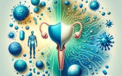
Leaky gut syndrome is really a nickname for the formal term “intestinal permeability” which is associated with many illnesses and symptoms. “It’s a symptom of inflammation and imbalance that has many causes” (Lipski, 2013). Leaky gut is associated with a variety of medical conditions such as allergies, celiac disease, inflammatory bowel disease (IBD), as well as many autoimmune diseases such as eczema, rheumatoid arthritis, Hashimoto’s thyroiditis, type 1 diabetes, systemic lupus erythematosus. I think in the next few years, Interstitial Cystitis (IC) will be added to the list as well. Below I will summarize what the role of the gut barrier is, some information about leaky gut syndrome, and demonstrate what the data infers on the association of leaky gut and IC. Although there are not any studies that have specifically analyzed the leaky gut theory among the IC population, I believe this is an area of great interest and can be monumental in revamping therapies aimed at addressing the root cause to facilitate healing and remission.
A summary of the intestinal barrier
Your gut lining is formally called the intestinal barrier, and often times termed the epithelial layer. It is a single-cell layer that constitutes the largest and most important barrier against the external environment. (Groschwitz & Hogan, 2009). It protects you from the external world of microbes, chemicals, toxins, etc. The intestinal barrier consists of various commensal microbiota that interact with the barrier to provide a protective function for the intestinal tissue. “The intestinal microbiota is considered to be largely symbiotic in nature and involved in various processes, including the breakdown and absorption of nutrients, production of vitamins and hormones, and prevention of colonization by pathogenic bacteria” (Bischoff et al., 2014). The function of the barrier is specialized, and it works to block the entry of diverse exterior antigens while having the ability to absorb key nutrients from your diet (Mu, Kirby, Reilly, & Luo, 2017).
When we talk about the intestinal barrier, we are mostly referring to the small intestine. However, the mucosal layer often can refer to a broader category that involves the mouth, through the stomach, intestines, colon and even the bladder. When we refer to “leaky gut”, it infers the gut is the “small intestine”. This is where all the magic happens. The small intestine is where most of the nutrient absorption occurs, where fats, proteins and starches can pass through and become assimilated (Lipski, 2013). In addition, the small intestine functions to keep bacterial products, foreign substances, and large digested molecules out, which is why it is referred to as a “barrier” (Lipski, 2013). The cells of the intestinal barrier form tight junctions, which are designed to not allow large molecules to pass through. Additionally, the cells also consist of a “brush border” which are just the layer of cells designed to allow the right nutrients and molecules to pass through. Therefore, the intestinal barrier wears two hats: Protection and Nourishment. What I mostly focus on is its protective role in this review, since many of the triggers of IC are related to bacterial overgrowth and infections.
According to Mu et. al, the gut barrier provides 3 main protective barrier functions: a physical barrier, a biochemical barrier and an immunological barrier. As a physical barrier, the intestinal lining consists of cells that are responsible for nutrient uptake as well as controlling the abundance of bacteria through expression of antimicrobial proteins (Mu et al., 2017). In addition, the gut consists of commensal bacteria that acts as a physical barrier that can promote resistance to colonization of pathogenic bacteria species by competing for nutrients, occupying attachment sites and releasing antimicrobial substances. Within the antimicrobial properties are the biochemical molecules that aid in reducing the load of colonized bacteria and are a good supplement with the physical barrier. And finally, the barrier consists of a variety of immune cells that regulate the immune response. A major role of the immunological barrier is secretory IgA (SIgA), the most abundant immunoglobulin in the body that resides primarily on intestinal mucosal surfaces (Mu et al., 2017). In fact, almost every lab I have viewed of clients with IC are low in SIgA, although a small percentage also demonstrate elevated levels.
Why is SIgA so important?
I would like to get a bit more “science geeky” and discuss the role of SIgA further, because I feel there is a strong link to this immunological barrier and the pathophysiology of IC. Throughout the digestive system we have immune tissue called GALT (gut-associated lymphoid tissue) and MALT (mucosal associated lymphoid tissue) that accounts for 70% of our immune system that protects us from antigens and foreign invaders (Lipski, 2013). SIgA is an abundant antibody that is produced and released by plasma cells that dominates humoral immunity at the intestinal mucosa (Chairatana & Nolan, 2017). “SIgA promotes the clearance of antigens and pathogenic microorganisms from the intestinal lumen by blocking their access to epithelial receptors, entrapping them in mucus, and facilitating their removal by peristaltic and mucociliary activities (Mantis et al., 2011). In fact, SIgA has a multi-faceted role in controlling inflammation and in regulating the immune responses to dietary antigens, commensal microflora and enteric pathogens (Mantis, Rol, & Corthesy, 2011). “When challenged by foreign molecules, SIgA forms immune complexes with allergens and microbes” (Lipski, 2013).
In just the past several years, SIgA’s role has been identified as the following (Mantis et al., 2011):
- It has the capacity to directly quench bacterial virulence factors
- It can influence the composition of intestinal microbiota
- It can promote retro transport of antigens across the intestinal epithelium
- It can downregulate proinflammatory responses normally associated with the uptake of highly pathogenic bacteria and allergenic antigens.
- It can combat microbial infections that are fundamentally different from those used by antibodies in systemic compartments
- It can interfere with the earliest steps in the infection process by blocking toxins and pathogens from literally adhering to the intestinal epithelium
From review of the most recent literature, it is becoming evident that altered levels of SIgA in the GI tract can contribute to many detrimental downstream effects, such as dysfunction in the immune recognition of intestinal bacteria, dysbiosis of the microflora (your gut bacteria), increased food allergies and food sensitivity issues (think gluten intolerance), more bacterial infections (think chronic UTI’s) and chronic low grade inflammation. SIgA can be elevated or lowered, depending on the condition of the patient. High levels of SIgA may indicate a variety of clues such as chronic stress, intestinal barrier dysfunction, acute GI dysfunction, among other things. Lowered SIgA levels, on the other hand, are associated with immune dysfunction and may indicate that the client has been under attack with infections for a very long time, and the body’s defenses have given up. All of these factors, by the way, can be associated with the presentation of interstitial cystitis as well.
What about Leaky Gut?
Leaky gut results from a damaged intestinal barrier. There are many causes of why this happens, the most common being chronic stress, dysbiosis (imbalanced bacteria), environmental toxins, GI disease, immune overload, poor food choices, pathogenic bacteria, parasites and yeast, and prolonged use of non-steroidal anti-inflammatory drugs (NSAIDs). Some of the clinical clues associated with leaky gut include abdominal bloating, gas, joint pain, maldigestion, mood swings, depression, poor immunity, recurrent bladder or vaginal infections, celiac disease or gluten intolerance, chronic fatigue syndrome, intestinal infections, IBS or IBD, psoriasis, eczema, and the list goes on and on.
Infections play a large hand in regulating the integrity of the mucosal barrier. A common example is a common bacteria known as H. pylori, a gram negative bacteria found in the human stomach. H. pylori can promote mechanisms that can increase leaky gut. In addition, microbial translocation (see next paragraph) can trigger an abnormal immune response, inflammation, and tissue damage in extraintestinal organs, and this includes the bladder!
What is even more interesting is the concept of “bacterial translocation”. This occurs when bacteria colonize in other parts of the body, which is often seen in leaky gut (Lipski, 2013). “For example, Bastocystis hominis, a parasite that causes GI problems, have been found in the synovial fluid of the knee of the arthritic patient” (Lipski, 2013). Certain microbiota, bacterial products, and metabolites affect the intestinal barrier function and are responsible for the subsequent breakdown of tissue homeostasis(Mu et al., 2017). “When there is a leaky gut, commensal bacteria in gut lumen, together with their products, are able to escape the lumen of the gut, which may induce inflammation and cause systemic tissue damages if translocated into peripheral circulation” (Mu et al., 2017). Theoretically, this concept can be applied to UTI’s and IC. Often times a GI stool analysis reveals that the same bacterial strains that are plaguing a patient’s bladder is also found overgrown in a stool test. I have seen this occur in may stool tests of IC patients that I have analyzed. A common bacteria that displays this mechanisms is E. coli. Factor in that many patients have low stomach acid, the conditions are perfect for translocation to occur in which the overgrowth of E. coli in the gut makes the person more susceptible to an E. coli infection in their bladder. I also find it interesting that many women with mycoplasma infections complain of having urethral pain. There are definitely some trends found in gut (as well as systemic) infections that needs to be explored deeper.
Be on the lookout for part to to learn about what other ways leaky gut can contribute to IC and bladder issues.
For more information about the Root Causes of IC, download the free Guide.
If you are ready to get started on the Root Cause investigation and start your healing journey, visit our website for more information and schedule a discovery call!
References
Bandara, H. M., Lam, O. L., Watt, R. M., Jin, L. J., & Samaranayake, L. P. (2010). Bacterial lipopolysaccharides variably modulate in vitro biofilm formation of Candida species. J Med Microbiol, 59(Pt 10), 1225-1234. doi:10.1099/jmm.0.021832-0
Bischoff, S. C., Barbara, G., Buurman, W., Ockhuizen, T., Schulzke, J. D., Serino, M., . . . Wells, J. M. (2014). Intestinal permeability–a new target for disease prevention and therapy. BMC Gastroenterol, 14, 189. doi:10.1186/s12876-014-0189-7
Burdette, Cheryl. (2017, December 20). LPS a Player in Chronic Disease. Retrieved (2018, April 16) from http://www.dunwoodylabs.com/index.php/webinar/
Chairatana, P., & Nolan, E. M. (2017). Defensins, lectins, mucins, and secretory immunoglobulin A: microbe-binding biomolecules that contribute to mucosal immunity in the human gut. Crit Rev Biochem Mol Biol, 52(1), 45-56. doi:10.1080/10409238.2016.1243654
Cui, Y., Liu, L., Dou, X., Wang, C., Zhang, W., Gao, K., . . . Wang, H. (2017). Lactobacillus reuteri ZJ617 maintains intestinal integrity via regulating tight junction, autophagy and apoptosis in mice challenged with lipopolysaccharide. Oncotarget, 8(44), 77489-77499. doi:10.18632/oncotarget.20536
de Punder, K., & Pruimboom, L. (2013). The dietary intake of wheat and other cereal grains and their role in inflammation. Nutrients, 5(3), 771-787. doi:10.3390/nu5030771
de Punder, K., & Pruimboom, L. (2015). Stress induces endotoxemia and low-grade inflammation by increasing barrier permeability. Front Immunol, 6, 223. doi:10.3389/fimmu.2015.00223
Groschwitz, K. R., & Hogan, S. P. (2009). Intestinal barrier function: molecular regulation and disease pathogenesis. J Allergy Clin Immunol, 124(1), 3-20; quiz 21-22. doi:10.1016/j.jaci.2009.05.038
Hong, H. A., Duc le, H., & Cutting, S. M. (2005). The use of bacterial spore formers as probiotics. FEMS Microbiol Rev, 29(4), 813-835. doi:10.1016/j.femsre.2004.12.001
Lee, B. J., & Bak, Y. T. (2011). Irritable bowel syndrome, gut microbiota and probiotics. J Neurogastroenterol Motil, 17(3), 252-266. doi:10.5056/jnm.2011.17.3.252
Lipski, E. (2013). Digestion Connection New York, NY: McGraw Hill.
Mantis, N. J., Rol, N., & Corthesy, B. (2011). Secretory IgA’s complex roles in immunity and mucosal homeostasis in the gut. Mucosal Immunol, 4(6), 603-611. doi:10.1038/mi.2011.41
McFarlin, B. K., Henning, A. L., Bowman, E. M., Gary, M. A., & Carbajal, K. M. (2017). Oral spore-based probiotic supplementation was associated with reduced incidence of post-prandial dietary endotoxin, triglycerides, and disease risk biomarkers. World J Gastrointest Pathophysiol, 8(3), 117-126. doi:10.4291/wjgp.v8.i3.117
Microbiome Medicine with Kiran Krishnan. (2017, April 21). Retrieved from https://www.youtube.com/watch?v=IolxUHdiX3Y&feature=youtu.be
Mu, Q., Kirby, J., Reilly, C. M., & Luo, X. M. (2017). Leaky Gut As a Danger Signal for Autoimmune Diseases. Front Immunol, 8, 598. doi:10.3389/fimmu.2017.00598
Ojetti, V., Petruzziello, C., Migneco, A., Gnarra, M., Gasbarrini, A., & Franceschi, F. (2017). Effect of Lactobacillus reuteri (DSM 17938) on methane production in patients affected by functional constipation: a retrospective study. Eur Rev Med Pharmacol Sci, 21(7), 1702-1708.
Turnbell, L., Mullin, Gerard, Weinstock, Leonard. (n.d.). Systemic Signs of Underlying Digestive Dysfunction and Disease. Principles of Integrative Gastroenterology.


")