

70% of the immune system in located in the gut (Lipski, 2013). The human body has several mechanisms that participate in regulating the immune system, including the innate immune system, adaptive immune system, immune-inflammatory response and regulatory system (Lipski, 2013). The innate immune system relies on certain receptors and secreted proteins that recognize common features of various pathogens (Chairatana & Nolan, 2017). The adaptive immune system employs allergic responses, are more specific to pathogenic invaders and take longer to respond to pathogens (Chairatana & Nolan, 2017).
The intestinal epithelium is a key contributor of innate immunity (Chairatana & Nolan, 2017). Throughout the digestive system we have immune tissue called GALT (gut-associated lymphoid tissue) and MALT (mucosal associated lymphoid tissue) that accounts for 70% of our immune system that protects us from antigens and foreign invaders (Lipski, 2013). “The intestinal epithelium is composed of four cell lineages, namely enterocytes, enteroendocrine cells, goblet cells, and Paneth cells, all of which are derived from a common stem cell progenitor in the intestinal crypts” (Chairatana & Nolan, 2017). The majority of epithelial cells bordering the intestinal lumen are absorptive enterocytes, which play a role in metabolism and digestion. Secretory intestinal epithelial cells (enteroendocrine cells, goblet cells, and Paneth cells) contribute to barrier function of the epithelium (Chairatana & Nolan, 2017). Cleary, mucosal immunity in the intestine involves a network of different types of cells that work together to provide barriers against microbes and also to maintain homeostasis (Chairatana & Nolan, 2017). If these surfaces are compromised, however, the bacteria, antigens and food particles can get into our blood and elicit inflammation and contribute to disease. As a result, defects in the innate immune system can result in increased susceptibility to Crohn’s disease and microbial infections (Chairatana & Nolan, 2017).
Secretory IgA (SIgA)
How does MALT convey the immune assault? Through a group of antibodies called Secretory IgA (SIgA). “The past several years have seen an emergence of evidence that indicates that SIgA influences the composition of the intestinal microbiota, promotes the uptake and delivery of antigens from the intestinal lumen to DC subsets located in GALTs, and influences inflammatory responses normally associated with the uptake of highly pathogenic bacteria and potentially allergenic antigens(Mantis et al., 2011).
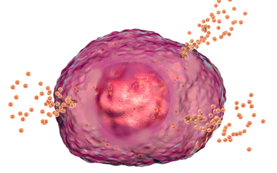
SigA is an abundant antibody that is produced and released by plasma cells that dominates humoral immunity at the intestinal mucosa (Chairatana & Nolan, 2017). Interestingly, it protects mucosal surfaces by non-covalently cross-linking microorganisms or macromolecules, and then promotes the clearance of the entrapped microbes by peristalsis or mucocilary movement (Chairatana & Nolan, 2017). This process is called immune exclusion: “SIgA promotes the clearance of antigens and pathogenic microorganisms from the intestinal lumen by blocking their access to epithelial receptors, entrapping them in mucus, and facilitating their removal by peristaltic and mucociliary activities (Mantis et al., 2011). In fact, SIgA has a multi-faceted role in controlling inflammation and in regulating the immune responses to dietary antigens, commensal microflora and enteric pathogens (Mantis et al., 2011). “When challenged by foreign molecules, SIgA forms immune complexes with allergens and microbes”(Lipski, 2013).
In just the past several years, SIgA’s role has been identified as the following (Mantis et al., 2011):
- It has the capacity to directly quench bacterial virulence factors
- It can influence the composition of intestinal microbiota
- It can promote retro transport of antigens across the intestinal epithelium to dendritic cell subsets in GALT
- It can downregulate proinflammatory responses normally associated with the uptake of highly pathogenic bacteria and allergenic antigens.
- It can combat microbial infections that are fundamentally different from those used by antibodies in systemic compartments
- It can interfere with the earliest steps in the infection process by blocking toxins and pathogens from literally adhering to the intestinal epithelium.
I also found it interesting that SIgA can coat commensal microorganisms in a newborn’s mucosal to favor gut colonization to educate their early immune system (Mantis et al., 2011). In all pathways, pathogenic and non-pathogenic bacteria are coated by SIgA. The interaction between SIgA-coated commensal bacteria and the epithelium reinforces its barrier function through multiple mechanisms, including reinforcement of tight junctions, overproduction of Polymeric immunoglobulin receptor( pIgR), and reduction in nuclear factor kappa-light-chain-enhancer of activated B cells nFk-B (Mantis et al., 2011). Note-pIgRs are mainly located on the epithelial lining of mucosal surfaces of gastrointestinal, respiratory, and genitourinary tracts where they are involved in the trancytosis of soluble IgA to the apical side of the mucosal epithelial (Polymeric immunoglobulin receptor, n.d.).
So how do you measure SIgA? There are labs that can run saliva tests. According to BioHealth labs, “measuring salivary levels of sIgA is a clinical biomarker that generally mirrors the sIgA levels in other mucosal tissues (e.g., gut) and has become a convenient tool to monitor changes in mucosal immune status” (BioHealth Lab, n.d.). Using the ELISA testing method, the lab uses saliva testing that can analyze SIgA levels over the course of a single day.
The current established reference ranges for sIgA (Biohealth Labs, n.d.)
Low: <75.0 μg/ml
Equivocal: 75.0-145.0 μg/ml
High: 145.0-330.0 μg/ml
Calprotectin and lactoferrin can also be measured to assess GI inflammation (Lipski, 2013). In fact, calprotectin and lactoferrin are often elevated in inflammatory bowel disease, postinfectious IBS, cancer in the digestive system, some GI infections, chronic infections, NSAID overuse, true food allergies and chronic pancreatitis (Lipski, 2013). Another marker of GI inflammation eosinophil protein X which can determine whether some has parasites, allergies, asthma or eczema (Lipski, 2013).
High levels of SIgA are associated with acute stress, intestinal barrier dysfunction, acute oral or GI infection, heavy smoking, alcoholism, periodontitis, dental plaque accumulation. In general, high levels point toward the need to rule out active infections in the body. It could also indicate malnutrition, malabsorption, or the lack of HCL production in the stomach, allergies, liver problems, parasites, or autoimmune conditions (Lipski, 2013).
Deficiency of SigA is the most common immunodeficiency, and low levels are associated with infections, autoimmune diseases, celiac disease, chronic infection, and food allergies (Lipski, 2013). “A study examining people with Crohn’s disease or ulcerative colitis found that all of them had low levels of SigA” (Lipski, 2013). So what can cause SIgA to become depleted? According to Lipski (2013), chronic stress, adrenal insufficiencies, oral bacteria, recurring infection, leaky gut, celiac disease, Crohn’s disease, viruses, immune hypersensitivity and anti-inflammatory drugs can all lower SIgA. Low levels may be an indication of autonomic nervous system imbalance, chronic stress, damage to the intestinal barrier, chronic GI infections, food intolerance, gliandin intolerance, IBD and/or use of NSAID’s or steroid medications (Biohealth, n.d.). Persistent low levels can lead to chronic infections in lung, gut or genitourinary tracts (Lipski, 2013).
In addition, overuse of antibiotics can contribute to modification of immune recognition of intestinal bacteria that can alter the levels of SIgA in the GI tract (Dzunkova et al., 2016). The study by Dzunkova et.al evaluated SIgA patterns in people who have C. difficile infections, which is often associated with broad-spectrum antibiotic use that can disturb normal gut microbiome. “The SIgA opsonization of commensals and pathogens results in two different outcomes: the first being that the commensal SIgA-coated bacteria are maintained within the gut lumen, and the second being that the SIgA-coated pathogens attempting to cross the epithelial barrier are removed” (Dzunkova et al., 2016). These findings of the study suggest that the growth of C. difficile may be suppressed under normal conditions by the beneficial bacteria found in the GI tract, while the decrease of their activity due to antibiotics induces a “blooming” of C. difficile (Dzunkova et al., 2016).
Lifestyle and nutritional factors can influence SIgA levels (Lipski, 2013). Interesting, studies indicate that raising SIgA might eliminate IBD (Lipski, 2013). Nutrients such as choline, essential fatty acids, glutathione, glycine, phosphatidylcholine, quercetin, vitamin C, and zinc are just some of the nutrients that are required to maintain healthy SIgA levels (Lipski, 2013).
Below are a list of things that can raise SIgA levels ((Lipski, 2013):
- Probiotics-saccharomyces boulardii, bifidobacteria longum, lactobacillus casei
- Conjugated linolenic acid
- Colostrum
- Transfer factor
- FOS
- MCT oil
- Rest, relaxation and singing (glad my daughter loves to sing!)
Another thing Liz mentions in her book is that rebalancing the gut is key to quenching the fire. An elimination diet can reduce inflammation and pain faster than anything else (Lipski, 2013). Eating a “rainbow” of polyphenols, such as cherries and berries, can also downregulate inflammation and protect blood vessels. Foods such as onions, low oxalate kale, broccoli, cabbage, low oxalate beans, a touch of celery, parsley, clover, as well as nutritional supplements such as ECGC (green tea), quercetin, curcumin, ginger, boswellia, bromelain, alpha lipoic acid, omega-3 fats, resveratrol and vitamin D (Lipski, 2013). This is why I really like the Microbiome mashup, it allows you to obtain these without going over too much in oxalate.
Another thing that was mentioned in the book for resetting the immune system was using TSO whipworm eggs, which can help people with chronic inflammation, oxidative stress, and poor detoxification. “Studies show dramatic remission rates in people with Crohn’s disease and ulcerative colitis as well as allergies, eczema, MS, asthma, hay fever, type I diabetes and food sensitivities” (Lipski, 2013). More research on my plate of things to look into!
To learn more about healing form IC naturally, please visit our website: https://ichealer.com
Instagram handle: drmandydcn
Threads username: drmandydcn
Tik Tok handle : drmandydcn
References
Biohealth Laboratory. (n.d.) Secretory IgA: The First Line of Immune Defense. Retrieved from https://www.biohealthlab.com/test-menu/immunity/.
Chairatana, P., & Nolan, E. M. (2017). Defensins, lectins, mucins, and secretory immunoglobulin A: microbe-binding biomolecules that contribute to mucosal immunity in the human gut. Crit Rev Biochem Mol Biol, 52(1), 45-56. doi:10.1080/10409238.2016.1243654
Dzunkova, M., Moya, A., Vazquez-Castellanos, J. F., Artacho, A., Chen, X., Kelly, C., & D’Auria, G. (2016). Active and Secretory IgA-Coated Bacterial Fractions Elucidate Dysbiosis in Clostridium difficile Infection. mSphere, 1(3). doi:10.1128/mSphere.00101-16
Lipski, E. (2013). Digestion Connection New York, NY: McGraw Hill.
Mantis, N. J., Rol, N., & Corthesy, B. (2011). Secretory IgA’s complex roles in immunity and mucosal homeostasis in the gut. Mucosal Immunol, 4(6), 603-611. doi:10.1038/mi.2011.41


")